CASE REPORT
A 57-year-old male patient was seen at our emergency department with flank pain and haematuria that had been present for three days. Nine months earlier he had received an allogeneic stem cell transplantation (alloSCT) for acute myeloid leukaemia. This alloSCT was complicated by a cytomegalovirus reactivation and a probable pulmonary invasive aspergillosis treated with valganciclovir and voriconazole, respectively. Five months after alloSCT he developed extensive chronic graft-versushost disease necessitating treatment with prednisolone and cyclosporine.
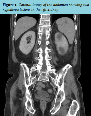
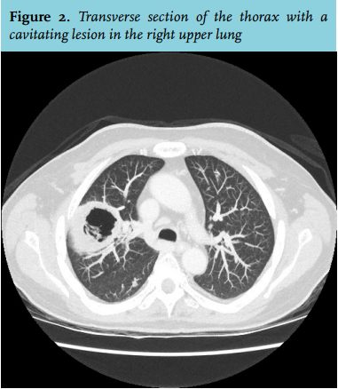
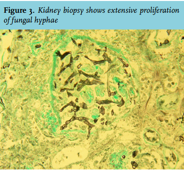
On the day of admission his medication consisted of prednisolone (40 mg once daily), cyclosporine (200 mg twice daily), valaciclovir (500 mg twice daily, prophylaxis for herpes virus reactivation), trimethoprimsulphamethoxazole (480 mg once daily, prophylaxis for Pneumocystis jiroveci pneumonia) and insulin because of steroid-induced diabetes mellitus. Physical examination revealed prominent left flank pain on percussion and palpation, but was otherwise unremarkable. The patient was afebrile and exhibited no pulmonary signs or symptoms. Laboratory data were as follows: leukocyte count 5.2 (normal range 4.0-11.0 x 10⁹/l) with a normal neutrophil count, haemoglobin 6.6 (normal range 8.4-10.8 mmol/l), platelet count 132,000 (normal range 150-400 x 109/l), serum creatinine 70 (normal range 60-110 µmol/l), serum glucose 8.0 mmol/l and C-reactive protein 204 (normal range < 10 mg/l). Urine analysis showed erythrocytosis and leukocytosis, without dysmorphic erythrocytes or red blood cell casts and the nitrate test was negative. An ultrasound was performed to evaluate the left kidney and surrounding organs and revealed a mass in the left kidney raising the suspicion of a renal cell carcinoma or lymphoproliferative disease. Therefore, a contrastenhanced computed tomography scan of the thorax and abdomen was performed which showed two masses in the left kidney and a cavitating lesion in the right upper lung (figure 1 and 2). A biopsy of the kidney (figure 3) and bronchoalveolar lavage were performed.
WHAT IS YOUR DIAGNOSIS?