Dear Editor,
Primary Sjögren syndrome (pSS) is a chronic autoimmune disease characterised by sicca syndrome. The diagnosis of SS relies on immunological and histological criteria,1 and salivary gland ultrasonography (SGUS) appears to contribute to diagnosis and follow up of pSS.2 The objective of our study was to assess the usefulness of SGUS in diagnosing SS. We performed SGUS for patients with pSS and controls suffering from sicca syndrome not fulfilling American-European Consensus Criteria for pSS.3 A positive SSA/SSB antibody test or a positive biopsy of minor salivary gland (focus score > 1) was requested to confirm pSS. Written informed consent was obtained from all patients.
The parotid and submandibular glands were examined by ultrasonography. The modified scoring system described by Cornec et al.4 was used to evaluate echogenicity and homogeneity for each gland. It ranged from grade 0 (normal gland) to grade 4 (multiples hypoechoic area or calcifications). A score > 2 was considered abnormal. 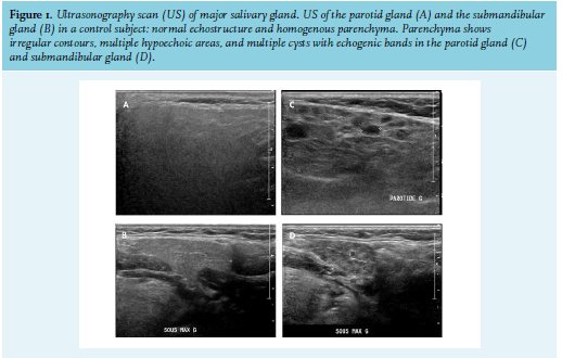
We enrolled 55 patients (30 pSS and 25 controls). In the pSS group, 22 patients had positive minor salivary gland (MSG) biopsy and 25 patients had positive SSA antibodies. Causes of sicca syndrome in the sicca group were diabetes (n = 5), idiopathic origin (n = 10), drug-induced (n = 4), sarcoidosis (n = 4), and hypothyroidism (n = 2). The two groups were comparable in terms of demographic characteristics. Twenty-one patients with pSS had an ultrasonography scan (US) score ≥ 2 and only three patients in the sicca group had a US score ≥ 2 (p = 0.001) (figure 1). The optimal cut-off for a US score was set at ≥ 2, with a sensitivity of 73% and specificity of 88%.The overall US score was correlated directly with SSA antibodies (r = 0.4, p = 0.002), MSG biopsy (r = 0.3, p = 0.002), and the diagnosis of pSS (r = 0.43, p = 0.001). Our study confirms the value of SGUS in contributing to a diagnosis of pSS. Our results suggest that SGUS could avoid MGS biopsy in 21 patients with pSS.
A group of experts suggested that SGUS had a similar weight to minor criteria of 2016 American College of Rheumatology/European League Against Rheumatism classification and improved sensitivity in diagnosing pSS.5 Other reports demonstrated that abnormal SGUS is associated with high disease activity and damage in pSS, suggesting that SGUS may be integrated in prognostic and therapeutic algorithms.6
Our study, although limited by the small number of cases, puts forward the usefulness of SGUS in diagnosing SS by detecting functional and structural impairments. SGUS abnormalities, in combination with clinical and immunological arguments, seems to be helpful for the diagnosis of SS.
DISCLOSURE
All authors declare no conflicts of interest. No funding or financial support was received.
REFERENCES