DIAGNOSIS
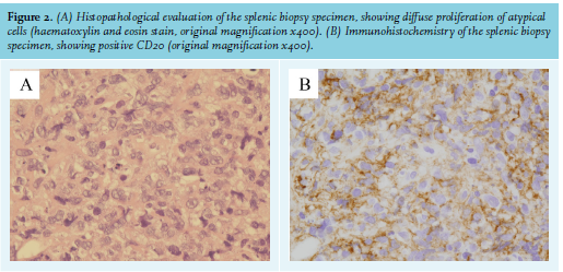
The differential diagnosis was angiosarcoma, splenic metastasis, or malignant lymphoma. Ultrasound-guided splenic core needle biopsy led to the diagnosis of diffuse large B-cell lymphoma (figure 2). Positron emission tomography-computed tomography indicated the presence of small lesions in the left scapula and the left ilium; thus the patient underwent chemotherapy with R-CHOP regimen. The patient is in complete remission.
Splenic incidental lesions are often encountered with abdominal CT. Although most of them are benign and are of no clinical significance, clinicians should pay closer attention to certain findings such as presence of solid, contrast-enhancing components and ill-defined borders.1 These warning findings indicate potentially more relevant diseases including malignancy, abscess, and sarcoidosis. Although splenectomy is generally used for diagnosis, fine needle biopsy and core biopsy are of considerable diagnostic value of suspicious splenic lesions.2
Lymphoma represents the most common malignant neoplasm of the spleen. While the spleen is usually involved secondarily in patients with lymphoma, isolated primary splenic lymphoma comprises less than 2% of all lymphomas. Lymphoma of the spleen may present on abdominal CT as splenomegaly without focal lesions, multiple miliary nodules, multiple lesions, or a single solitary mass.3 Although lymphomatous involvement of the spleen is seldom an incidental finding, it may be seen in patients with vague symptoms including left upper abdominal pain, weight loss, and fatigue. Because early recognition and immediate treatment are crucial for patient prognosis, clinicians should consider lymphoma as a possible differential diagnosis of splenic incidentaloma, especially with warning findings.
DISCLOSURE
All authors declare no conflicts of interest. No funding or financial support was received.
REFERENCES