KEYWORDS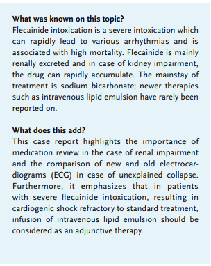
Cardiogenic shock, flecainide intoxication, intralipid, intravenous lipid emulsion, sodium bicarbonate.
INTRODUCTION
Flecainide acetate (Tambocor) is a Vaughn-Williams class 1C anti arrhythmic drug and a sodium and potassium channel blocker, mainly used for the treatment of supraventricular arrhythmias but also for ventricular dysrhythmias.1,2 The therapeutic window for flecainide is narrow and 80-90% of it is eliminated (of which 25-40% is unchanged) by the kidneys.3 Severe intoxication is associated with a mortality rate of approximately 10%.4 Here, we present a patient with a severe flecainide intoxication on a background of kidney impairment, who was admitted at the Intensive Care Unit (ICU) and successfully managed with the infusion of sodium bicarbonate and intravenous lipid emulsion (ILE). This case illustrates the importance of prompt symptom recognition and the recognition of medication as the cause of a collapse, especially in the case of kidney dysfunction. Furthermore, it highlights the importance of accurate comparison of new and old ECGs.
CASE REPORT
A 68-year-old female patient with a medical history of a sick sinus syndrome, for which a dual-chamber, rate-modulated pacemaker was implanted, atrial fibrillation, and chronic renal failure, presented to the emergency department after collapsing. Her home medication consisted of bisoprolol, bumetanide, flecainide and rivaroxaban. Except for bumetanide, this medication was continued at admission.
An old ECG is shown in figure 1 and the ECG at the time of admission to the emergency department is shown in figure 2. Ischemia was ruled out since troponin was negative and a recently performed coronary angiography was normal. Her pacemaker report did not reveal arrhythmias. A urinary tract infection was diagnosed and further lab results revealed acute on chronic kidney failure, with a glomerular filtration rate (GFR) declining from 20 to 12 ml/min/1.73m2 and a creatinine rise from 219 to 318 µmol/l. Thus, intravascular fluid depletion due to the infection was deemed to be the cause of her collapse. 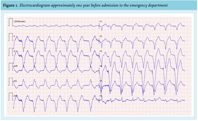
Several days later, she deteriorated with hypotension (inter-beat (RR) interval 80/45 mmHg), bradycardia (heart rate 45-50/min), somnolence, peripheral cyanosis and mottled extremities. She was admitted to intensive care.
Further work up revealed:
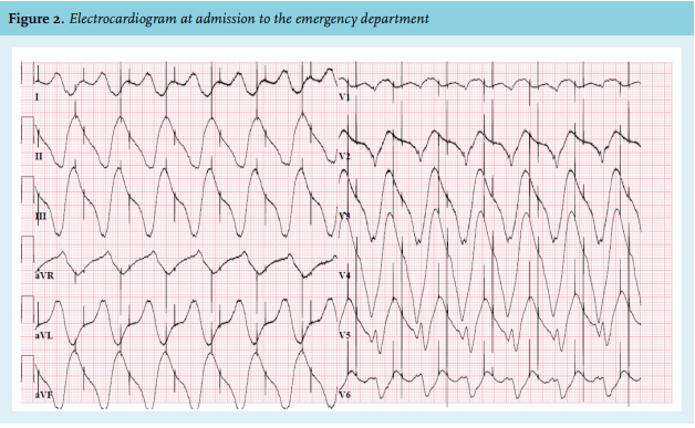
A new ECG (figure 3) demonstrated extreme broad QRS complexes and loss of pacemaker capture. A review of her medication showed that flecainide 200 mg had been continuously administered throughout her admission. We concluded that she was suffering from cardiogenic shock thought to be caused by flecainide toxicity. A flecainide level was obtained, which was indeed within the toxic range: 2.44 mg/l. Although deemed less likely, since our patient was only treated with a very low dose of bisoprolol (2.5 mg once daily) whilst in the coronary care unit (CCU), the contribution of bisoprolol toxicity to the symptoms described above could not be completely ruled out. For this reason, we also obtained bisoprolol levels, which were 0.10 mg/l at ICU admission and non-detectable the day after. Thus, we decided to consider the case as a mono-intoxication with flecainide.
The patient was treated with sodium bicarbonate infusion and intubated shortly after ICU admission because of respiratory exhaustion. She remained anuric and was started on continuous venovenous haemofiltration (CVVH). Despite further supportive treatment with noradrenaline, isoprenaline and dobutamine, hypotension and bradycardia persisted and pulse contour cardiac output measurements remained consistent with low cardiac output (cardiac index 1.4, extravascular lung water index 17.6, global end-diastolic volume index 756). Finally, intralipid 20% was administered at the maximum dosage, resulting in a gradual overall improvement. After 10 days, she was extubated and after 12 days discharged to the CCU.
DISCUSSION
This case report describes a patient who developed a flecainide intoxication, a rare but often fatal intoxication, because of continuous administration of the drug despite declining kidney function.
Flecainide’s main effects are exerted through its high affinity for open-state sodium channels, thereby prolonging the depolarisation of myocardial cells. This results in a reduction of cardiac excitability in all parts of the heart.1,2 The majority of flecainide is renally excreted, 80-90%, of which 25-40% unchanged, with a half-life of approximately 20 hours. At higher doses, flecainide demonstrates non-linear pharmacokinetics, which means elimination half-life increases at higher plasma levels. In severe kidney failure, elimination can be extended to as long as 58 hours and the drug can rapidly accumulate. The normal therapeutic range is 0.2 to 1 mcg/ml, although adverse effects have been reported with plasma levels > 0.7 mcg/ml.5,6 Our patient’s flecainide dose was not adjusted to her kidney function and flecainide levels were not monitored. This case report highlights the importance of medication review in case of kidney failure.
The clinical features of flecainide toxicity vary from blurred vision to hypotension and bradyarrhythmias as well as tachyarrhythmias. Severe intoxication can be fatal: the mortality rate of flecainide intoxication has been estimated at 10%.4,7 The electrophysiologic properties of flecainide are manifested on ECGs as PR and QTc interval prolongation and QRS widening.8,9 When the QRS interval is widened more than 25% compared to baseline, this is considered as the threshold for dose reduction or discontinuation of the drug. When the QRS duration is increased by 50% or the PR interval is prolonged by 30%, toxicity should be suspected.8,10 In hindsight, when comparing ECG 1 and 2, we can conclude that QRS complexes were already profoundly wider at admission to the emergency department. This should have prompted the treating clinicians to consider flecainide as the cause of her collapse and reduce the dosage or discontinue the drug.
Since flecainide intoxication is rare, literature on treatment options is limited.7 In addition to ICU admission for invasive supportive management and correcting aggravating conditions for arrhythmias, such as hypoxia and electrolyte disturbances, high-dose sodium bicarbonate infusion is the cornerstone of treatment. It acts by antagonizing flecainide at its binding site to sodium channels on cardiac myocytes. Aggressive treatment is required and doses up to 350 mEq have been reported to maintain a goal pH of 7.45-7.55.6,11 Because of the refractory state of the cardiogenic shock in this patient, ILE was also administered. This is a novel strategy and has only been described in a few case reports.12-14 The mechanism of action is not completely understood, but various potential mechanisms have been proposed. The most prevalent is the ‘lipid sink’ theory, suggesting that with lipid administration, the intravascular lipid phase is expanded, which extracts the offending lipid soluble drug from its target tissue. Another hypothesis theorises that lipid administration serves as a myocardial energy substrate, thereby improving cardiac function.15 Our patient was successfully managed with ILE and we think it should be considered as adjunctive therapy in patients with refractory cardiogenic shock secondary to flecainide overdose.
DISCLOSURES
All authors declare no conflicts of interest. No funding or financial support was received.
REFERENCES