CASE REPORT
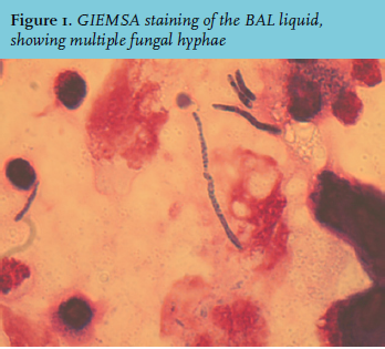
A 67-year-old woman was admitted to the hospital with progressive dyspnoea during the last three weeks. Medical history revealed seronegative polyarthritis treated with methotrexate, colon carcinoma and extended hemicolectomy, diabetes mellitus type 2 and hypertension for which she was taking medication. She had been a smoker in the past and drank alcohol daily. Three months prior to admission the polyarthritis worsened, and prednisolone 15 mg daily was added. X-ray of the chest showed diffuse interstitial density, CT scan of the chest showed an atypical image of pronounced density and ground glass opacity. She was treated with penicillin, ciprofloxacin and furosemide, but did not improve. The pulmonologist suspected an immunocompromised state due to long-term use of methotrexate and prednisolone, and feared opportunistic Pneumocystis jiroveci pneumonia (PJP). A bronchoalveolar lavage (BAL) was performed and co-trimoxazole and high-dosed prednisolone was started. After the BAL her condition worsened and she was admitted to the intensive care unit (ICU). Physical examination showed a respiratory rate of 35/minute, heart rate of 98/minute, blood pressure of 150/80 mmHg and a tympanic temperature of 37.9°C. Blood gas analysis showed poor oxygenation (pH 7.45, pCO2 4.2 kPa, pO2 6.6 kPa, HCO3- 22.2 mmol/l, base excess -1.6 mmol/l, SatO2 85%) on high-flow nasal cannula oxygen therapy (Optiflow®; FiO2 = 100%, O2 -flow = 50 litres/minute). She had to be intubated and mechanically ventilated. Laboratory results showed a normal leukocyte and neutrophil count, C-reactive protein 98 mmol/l, and elevated lactic dehydrogenase 433 U/l. The diagnosis PJP unfortunately remained questionable; microscopic examination by silver staining and polymerase chain reaction of the BAL liquid did not show evidence for PJP, however the GIEMSA staining was dubiously positive. But, more surprisingly, fungal hyphae were also noticed (figure 1).
WHAT IS YOUR DIAGNOSIS?