Full textPDF
Full text
CASE REPORT
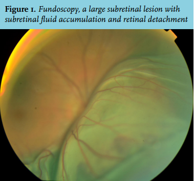
A 19-year-old Caucasian male with no previous medical history, presented to our outpatient clinic with a one-week history of pain, redness and decreased visual acuity (1/60) of the right eye. Fundoscopy showed a large subretinal lesion with subretinal fluid accumulation (figure 1). On further physical examination an elastic swelling with a diameter of 10 mm was found on his back. He had also had an enlarged right testicle for as long as he could remember, and he had not noticed a difference in the last five years. Initial laboratory investigations showed a slightly elevated lactate dehydrogenase (282 IU/l). B-scan ultrasonography of the eye revealed an irregular, medium-high reflective choroidal tumour with a diameter of 20 mm and a thickness of at least 12 mm.
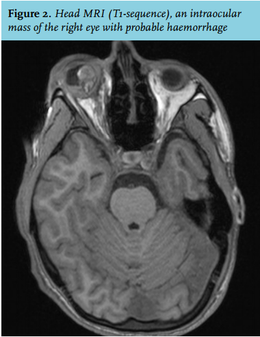
Computed tomography of chest and abdomen showed multiple intra-pulmonary lesions, suspected for metastases. Magnetic resonance imaging of the brain Figure 1. Fundoscopy, a large subretinal lesion with subretinal fluid accumulation and retinal detachment Figure 2. Head MRI (T1-sequence), an intraocular mass of the right eye with probable haemorrhage showed an intraocular mass of the right eye with haemorrhage (figure 2) and an intracerebral lesion.
WHAT IS YOUR DIAGNOSIS?