CASE REPORT
A 34-year-old, otherwise healthy man visited the emergency department with sudden painless paralysis of both legs for the past three days. Apart from trouble with urinating and feeling feverish, he had no other symptoms. When asked, the patient admitted to excessive alcohol and nicotine use of up to 3 litres beer and 6-12 cigarettes a day. He declined using drugs.
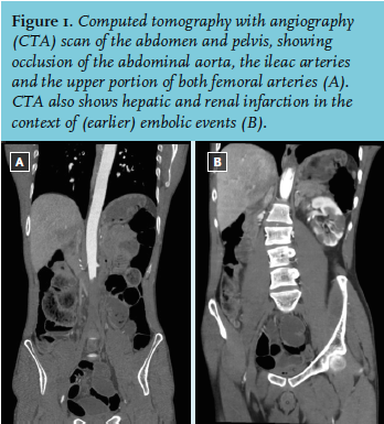
On physical examination, an Afro-American male was seen. He had a pulse of 123 beats/min, his blood pressure was 154/108 mmHg. His temperature was 35.7° Celsius, and his SpO2 was 97%. He had a complete paralysis of both his legs, and loss of sensibility. Both his legs were pale and cold. The pulsations of the femoral, popliteal, anterior tibial and dorsalis pedis arteries were absent on both sides. The neurological examination was normal: motoric and sensory function of the cranial nerves was intact, there was no paralysis or loss of sensibility of his upper extremities and the coordination was not impaired. The electrocardiogram (ECG) showed a sinus tachycardia of 130 beats/min, but no other abnormalities. Laboratory tests revealed a slightly elevated leukocyte count of 17.1 (normal value: 4.0-10.0 x 10⁹/l), a C-reactive protein of 94 (CRP: 0-8 mg/l), an elevated lactate dehydrogenase level of 3068 (LDH: 0-248 U/l), a creatinine kinase level of 128,932 (CK: 0-145 U/l), an aspartate transaminase of 2155 (ASAT: 0-31 U/l), and an alanine transaminase of 365 (ALAT: 0-34 U/l). The arterial blood sample was normal with the exception of an elevated lactate of 2.9 (0.5-1.7 mmol/l). His vitamin B1, B6, B12 and his folic acid levels were within the normal range. Lumbar puncture showed a leukocyte count of 1.0 (0.0-5.0 x 10⁶/l), a glucose of 4.2, and an albumin of 115.9 (100-300 mg/l); there were no erythrocytes. Magnetic resonance imaging of the cerebrum, and thoracic and lumbar spine showed no abnormalities. A computed tomography with angiography scan was performed (figure 1).
WHAT IS YOUR DIAGNOSIS?