CASE REPORT
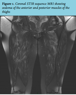
A 44-year-old woman presented with pain in her arms and legs and walking difficulties lasting for one month, partially relieved by ketoprofen. She had a medical history of primary Sjögren syndrome (PSS) (5/6 criteria of the American-European Consensus Group were fulfilled: ocular and oral symptoms, ocular signs, lymphocytic sialoadenitis with a focus score ≥ 1 on minor labial accessory gland, and positive anti-SSA antibodies) and autoimmune thyroiditis, and was treated with hydroxychloroquine and levothyroxine. On physical examination, she appeared healthy, her temperature was 37.8°C. She could move her arms but was unable to walk because of thigh pain and weakness. The remainder of the physical examination was normal. Her young son had had a febrile rash six weeks before. The blood cell count, serum creatinine, creatinine phosphokinase, lactate dehydrogenase, thyroidstimulating hormone, and urinalysis were unremarkable, the alanine aminotransferase level was 110 U/l (N < 32), aspartate aminotransferase 57 U/l (N < 32), C-reactive protein 79 mg/l (N < 5), serum electrophoresis showed an increased level of alpha-2 globulin and polyclonal hypergammaglobulinaemia. Antinuclear and anti-ECT antibodies titres were unchanged; a search for myositisspecific and anti-neutrophil cytoplasmic antibodies was negative. No cryoglobulinaemia was found, complement proteins C3 and C4 levels were increased. High levels of Parvovirus B19 (PvB19) immunoglobulin G (IgG) and immunoglobulin M (IgM) were found in her serum and the PCR was positive at 21,816 IU/ml. Electromyogram, thoraco-abdomino-pelvic scan and 18-fluorodeoxyglucose PET/computed tomography detected no abnormalities. MRI of the thighs showed diffuse muscular oedema without amyotrophia of the anterior and posterior muscles of the thighs (figure 1).
WHAT IS YOUR DIAGNOSIS?