Full textPDF
Full text
CASE REPORT
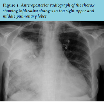
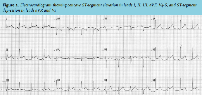
A 62-year-old woman was admitted to the emergency department because of severe fatigue, coughing, and fever. Her blood pressure was 90/65 mmHg, pulse frequency 108 beats/min, oxygen saturation 99%, and body temperature 38.5 °C. Pulmonary auscultation revealed bilateral rhonchi. Blood examination showed leukocytes of 16.3 x 10⁹/l, predominantly segmented granulocytes, a C-reactive protein concentration of 288 mg/l, renal insufficiency, anaemia, hypoalbuminaemia, raised liver enzyme concentrations, and a lactate concentration of 4 mmol/l. Radiography of the thorax showed infiltrative changes in the right upper and middle lung lobes, with a rounded heart contour (figure 1). A routinely obtained electrocardiogram showed concave ST-segment elevation in leads I, II, III, aVF, V4-6, and ST-segment depression in leads aVR and V1 (figure 2).
WHAT IS YOUR DIAGNOSIS?